

A Novel Imaging Study to Quantify Respiratory Function in Early Onset Scoliosis -
Introducing Quantitative Dynamic Magnetic Resonance Imaging (QdMRI1)
Yubing Tong, PhD; Jayaram K. Udupa, PhD; Joseph M. McDonough, MS; Caiyun Wu, MS; Catherine Qiu, MS; Carina Lott, MS; Nirupa Galagedera, BA; Jason B. Anari, MD; Drew A. Torigian, MA; Patrick J. Cahill, MD
Some children with scoliosis early in life can develop lung problems. The worst form is Thoracic Insufficiency Syndrome. This is where the lungs are too small. They also don’t move enough air in and out with each breath. Surgery for early onset scoliosis is done in hopes of improving the child’s breathing. Unfortunately, these results are hard to measure. The old-fashioned way to measure lung function is spirometry. In this test, the patient is required to breath as deeply as possible into a test device. Unfortunately, young children can’t perform this test well. The goal of this study was to develop a new way to measure lung function that doesn’t count on a young child’s effort.
Cahill and colleagues from the Children’s Hospital of Philadelphia came up with a new way to measure lung function in these children. They use magnetic resonance imaging (MRI) to look at the lung during breathing. An MRI can see soft tissues such as lung and measure their size precisely. They measure the change from breathing in to breathing out. They can then measure important features such as how big a breath the patient can take and how much air moves in and out with each breath. Even better, they can tell how well each of the lungs works. They can also tell whether the rib cage or the breathing muscles help more.
The authors have looked at a group of 30 kids who had surgery. The surgery was done because of severe scoliosis or breathing problems. Each had a titanium rib device placed. This is also called a VEPTR (Vertical Expandable Prosthetic Titanium Rib). It is an internal device that attaches to the ribs to hold them up. The VEPTR can be enlarged with a small surgery as the child grows. Their MRI method was used to measure lung function.
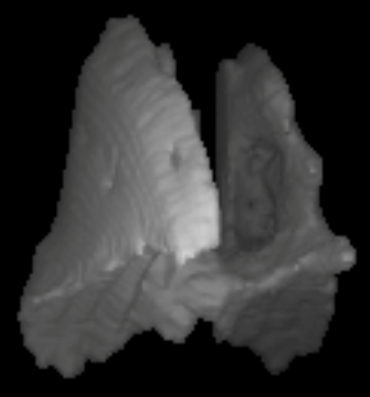
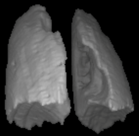
They found that after surgery, the lungs enlarged about thirty percent better. Further, the amount of air moving with each breath improved by over 50 percent. The right-side lung was often worse before surgery. It improved more with surgery, though. The figure below shows a picture of the lungs before and after surgery with this test. Kids can take a deeper breath after surgery.
| Before Surgery | After Surgery |
 |
 |
They also showed that their measurements were accurate. They did this by comparing their measures to the old-fashioned test. This new method shows promise in further understanding how lungs are helped by this surgery.
Summary provided by the SRS Patient Education Committee